
Dangerous Chicken Bone Lodged in the Esophagus — Removed Without Open-Chest Surgery
Esophageal foreign bodies are among the most dangerous emergency conditions in dogs, especially when sharp objects become lodged deep inside the chest cavity. In many cases, thoracic surgery is considered the standard treatment. This case demonstrates how a portable veterinary endoscope enabled the successful removal of a sharp chicken fork bone from the thoracic esophagus — avoiding open-chest surgery entirely.
Case Source
Luoyang Aita Animal Hospital, Henan
Case Background
- Species: Dog
- Gender: Male
Chief Complaint
The dog accidentally swallowed a sharp chicken fork bone that became lodged in the thoracic esophagus near the cardiac region.
Due to the dangerous location and sharp structure of the foreign body, thoracic surgery was widely considered necessary.

Clinical Challenge
Sharp esophageal foreign bodies present major clinical risks:
- Esophageal perforation
- Bleeding
- Severe infection
- Mediastinal complications
- Difficulty of surgical access
Traditional open-chest surgery is highly invasive and often requires prolonged recovery.
Radiographic Examination
X-ray imaging was first performed to confirm the exact location of the foreign body.
- Sharp foreign object identified in the thoracic esophagus
- Location confirmed near the cardiac region

Endoscopic Procedure
The veterinary team performed esophageal endoscopy using the JeetVet RAE-105Pro portable veterinary endoscope.
- Endoscope inserted through the oral cavity into the esophagus
- Continuous air insufflation for clear visualization
- Precise localization of the chicken fork bone
- Foreign body forceps inserted through the instrument channel
- Bone carefully grasped and removed through the mouth

Key Findings
- Sharp chicken fork bone lodged in the thoracic esophagus
- Localized mucosal irritation
- No severe perforation detected
The foreign body was successfully removed without the need for thoracic surgery.
Treatment Outcome
- Rapid recovery after anesthesia
- No chest incision required
- Minimal postoperative discomfort
Remarkably, the dog regained normal activity within just 10 minutes after the procedure.

Endoscopic Removal vs Open-Chest Surgery
| Comparison | JeetVet Endoscopic Removal | Open-Chest Surgery |
|---|---|---|
| Trauma | Minimally invasive, almost no external incision | Major thoracic incision |
| Anesthesia | Short sedation/general anesthesia | Long full anesthesia |
| Recovery Speed | Fast, often same-day discharge | Slow, hospitalization for 1–2 weeks |
| Complication Risk | Low | Higher risk of infection and cardiopulmonary complications |
| Pain Level | Mild | Significant |
| Cost | Lower | Higher |
Why Endoscopy Was Critical
- Minimally Invasive: Avoided traumatic thoracic surgery
- Precise Visualization: Enabled safe extraction of the sharp foreign body
- Faster Recovery: Reduced pain and shortened recovery time
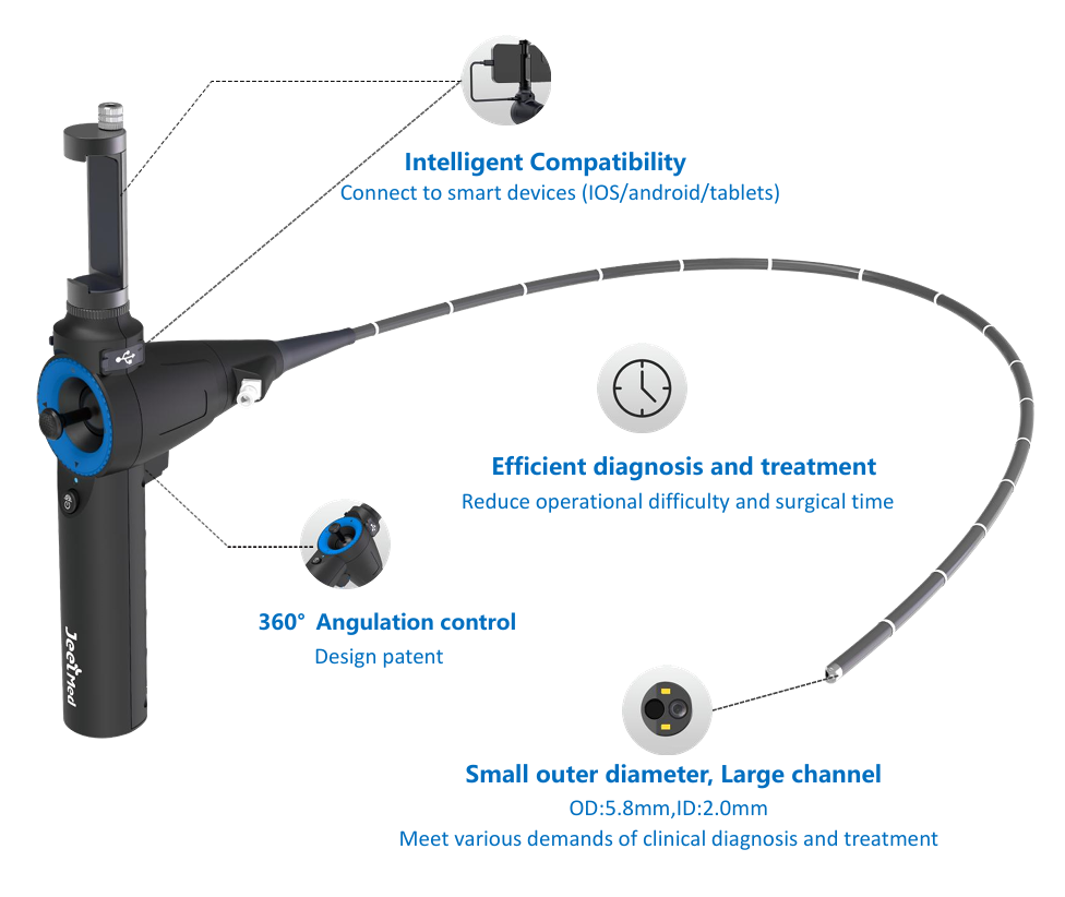
JeetVet Endoscope Advantage
JeetVet RAE-105Pro portable veterinary endoscope is designed for difficult foreign body procedures:
- 5.8 mm slim insertion tube
- 360° articulation for flexible maneuvering
- 2.0 mm working channel for retrieval tools
- Portable connection to phone and computer
- Suitable for gastrointestinal and esophageal applications
Clinical Value
Compared with traditional thoracic surgery, veterinary endoscopy offers:
- Reduced trauma
- Lower complication risk
- Faster recovery
- Improved patient comfort
Conclusion
Sharp esophageal foreign bodies can rapidly become life-threatening if not treated promptly.
A portable veterinary endoscope provides a safe, minimally invasive solution for complex foreign body removal, helping avoid open-chest surgery and dramatically improving recovery outcomes.

