
Dog Swallowed a Sock? See How Endoscopy Solved It Without Surgery
Foreign body ingestion is a common emergency in small animal practice. This case demonstrates how a portable veterinary endoscope enabled a fast, safe, and minimally invasive solution.
Case Source
Henan Aita Animal Hospital
Patient Background
- Breed: West Highland White Terrier
- Gender: Male
- Age: 3 months
Clinical Symptoms
The puppy accidentally swallowed a sock, leading to severe vomiting. Despite the symptoms, the dog remained in relatively stable condition with no signs of systemic infection.

Procedure Overview
The veterinary team first used X-ray imaging to locate the foreign object. The dog was then placed under general anesthesia.
A JeetVet RAE-105P veterinary endoscope was inserted through the oral cavity. Using the JeetVet irrigation and suction pump, the stomach was insufflated to improve visibility.
Once the sock was identified, forceps were introduced through the working channel to successfully remove it.


Core Surgical Approach
Under full anesthesia, the endoscope was guided from the mouth through the esophagus into the stomach to retrieve the swallowed sock.
This method is ideal when the foreign object is still located in the stomach, significantly reducing surgical risks.
Key Procedure Steps
- Fasting and fluid therapy to correct dehydration and electrolyte imbalance
- X-ray or ultrasound to confirm foreign body location
- Pre-anesthetic blood tests (CBC, biochemistry, coagulation)
- Stable anesthesia with intubation to prevent aspiration
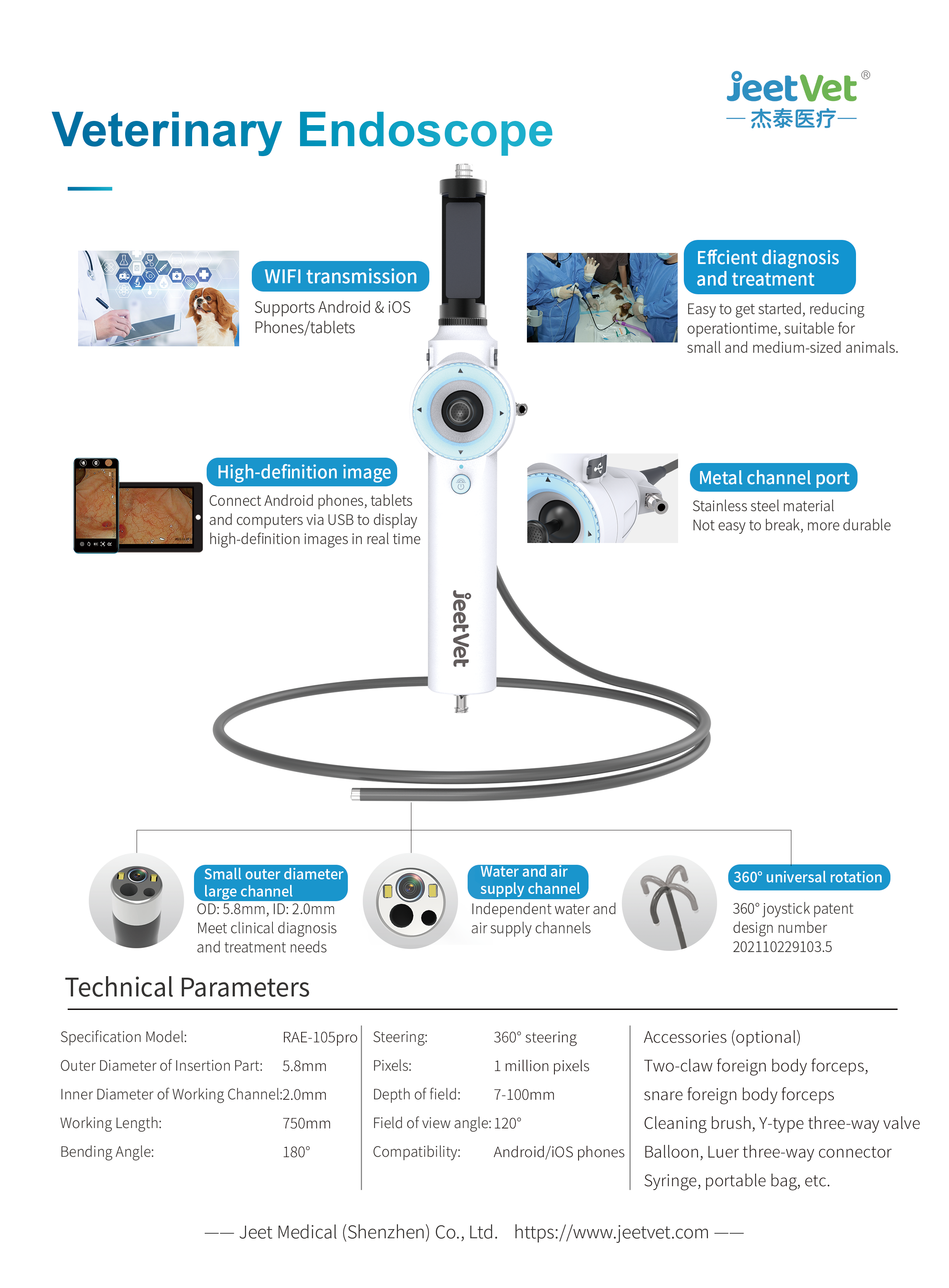
- Insertion of 5.8mm diameter / 2.0mm channel endoscope (RAE-105P)
- Use of irrigation pump for controlled insufflation (4–6 mmHg)
- Foreign body removal using endoscopic forceps
Advantages vs Traditional Surgery
| Aspect | Endoscopic Removal | Open Surgery |
|---|---|---|
| Invasiveness | Minimally invasive | Highly invasive |
| Recovery | Fast recovery | Long recovery time |
| Pain Level | Minimal | High |
| Procedure Time | Short | Long |
| Cost | Lower | Higher |
Limitations
- Foreign body located in the distal intestine
- Severe impaction or intestinal necrosis
- Perforation or peritonitis
- Sharp or rigid objects (bones, needles)
- Failure to grasp object after multiple attempts
Postoperative Care
- Fasting: 12–24 hours if no mucosal damage
- Medication: Antiemetics and gastric protectants
- Fluids: Correct dehydration and electrolytes
- Monitoring: Observe vomiting, appetite, and activity
- Diet: Soft, digestible food in small portions
Why Choose JeetVet Endoscope?
This case highlights the clinical value of JeetVet portable veterinary endoscope systems:
- High-definition imaging for accurate diagnosis
- Dual-channel design for treatment + visualization
- Portable and easy to operate
- Ideal for small animal clinics and emergency cases
Conclusion
Endoscopic foreign body removal is a safe, efficient, and minimally invasive solution. Compared to traditional surgery, it significantly improves recovery time and reduces risk.
For clinics and distributors, investing in a portable veterinary endoscope is a strategic move toward advanced veterinary care.

