Veterinary Endoscope for Cats: How Gastroscopy Helps Investigate Chronic Vomiting
Chronic vomiting is a common but sometimes challenging clinical problem in cats. Occasional vomiting may be associated with dietary changes or hairballs, but repeated or persistent episodes can indicate underlying gastrointestinal disease that requires further investigation.
When routine examinations cannot fully explain the symptoms, a veterinary endoscope can provide veterinarians with a direct view of the esophagus, stomach, and accessible portions of the upper gastrointestinal tract.
More importantly, veterinary gastroscopy can allow targeted tissue samples to be collected from abnormal or clinically relevant areas, helping clinicians move from visual assessment toward a more definitive diagnosis.
When Does Chronic Vomiting Require Further Investigation?
Vomiting is a clinical sign rather than a diagnosis. When it becomes frequent, persistent, or is accompanied by other abnormalities, further diagnostic evaluation may be appropriate.
Veterinarians may be particularly concerned when vomiting occurs together with:
- Reduced appetite
- Progressive weight loss
- Lethargy
- Repeated regurgitation or gagging
- Blood in vomit
- Changes in stool quality
- Failure to respond to initial treatment
The diagnostic approach depends on the individual patient and may include laboratory testing, diagnostic imaging, and, when clinically indicated, gastrointestinal endoscopy.
Why Use a Veterinary Endoscope?
A veterinary endoscope provides something many other diagnostic approaches cannot: direct visualization of the gastrointestinal mucosal surface.
During gastroscopy, veterinarians may evaluate:
- Mucosal color and texture
- Inflammation
- Erosions and ulceration
- Abnormal tissue or masses
- Foreign material
- Changes around the pyloric region
Visual findings alone, however, do not always establish a definitive diagnosis. This is why biopsy capability is an important part of gastrointestinal veterinary endoscopy.
What Happens During Feline Gastroscopy?
Upper gastrointestinal endoscopy is generally performed under general anesthesia. The patient is prepared according to the veterinary team's protocol, including appropriate fasting and pre-anesthetic evaluation.
A flexible veterinary endoscope is carefully introduced through the mouth and advanced through the esophagus toward the stomach.
The clinician can systematically examine areas including:
- Esophagus
- Gastroesophageal junction
- Gastric body
- Gastric fundus
- Antrum
- Pylorus
- Accessible proximal duodenum, depending on the patient and equipment
Careful navigation and adequate visualization are particularly important in feline patients because of their smaller anatomy.
The Importance of Endoscopic Biopsy
One of the major clinical advantages of a veterinary endoscope with an instrument channel is the ability to pass biopsy forceps through the scope while maintaining direct visualization.
This enables veterinarians to collect mucosal samples from selected locations without performing an open abdominal procedure.
Endoscopic biopsy may contribute to the investigation of conditions such as:
- Chronic gastritis
- Inflammatory gastrointestinal disease
- Selected infiltrative disorders
- Suspicious mucosal lesions
- Some gastrointestinal neoplasms
Collected samples can then be submitted for histopathological evaluation. Final diagnosis should be based on the complete clinical picture, including history, examination findings, laboratory results, imaging, endoscopic appearance, and pathology when appropriate.
Why Endoscope Diameter Matters in Cats
Cats present different anatomical requirements from medium and large dogs. For this reason, insertion tube diameter is an important consideration when selecting a veterinary endoscope for cats.
An appropriately selected flexible endoscope can help provide:
- Better access through smaller anatomical passages
- Controlled maneuverability
- Adequate visualization
- Reduced unnecessary mechanical pressure
- Access to a working channel when biopsy or intervention is required
The ideal configuration depends on patient size, intended procedure, working channel requirements, and the veterinarian's clinical assessment.
Gastroscopy Is More Than Looking Inside the Stomach
A common misconception is that endoscopy is simply a camera examination. In practice, a modern veterinary endoscope can serve as both a visualization platform and an access route for diagnostic or therapeutic instruments.
| Capability | Clinical Purpose |
|---|---|
| Direct Visualization | Assess the appearance of the gastrointestinal mucosa |
| Targeted Biopsy | Collect tissue samples from selected locations |
| Foreign Body Retrieval | Remove suitable objects when clinically appropriate |
| Image Documentation | Record findings for case review and follow-up |
| Post-Treatment Evaluation | Reassess selected lesions when clinically indicated |
Endoscopy Does Not Replace Every Diagnostic Method
Veterinary endoscopy should be viewed as part of a complete diagnostic workflow rather than a universal solution for chronic vomiting.
Some gastrointestinal diseases involve deeper layers of the intestinal wall or regions that cannot be adequately reached using routine upper gastrointestinal endoscopy. In these situations, additional diagnostic procedures may still be required.
The decision to perform endoscopy should therefore be based on the patient's clinical signs, previous diagnostic findings, and the veterinarian's judgment.
What Should Clinics Look for in a Veterinary Endoscope for Cats?
For clinics planning to expand feline gastrointestinal endoscopy, several equipment characteristics deserve attention:
- Appropriate insertion tube diameter
- Clear, stable image quality
- Reliable illumination
- Responsive tip articulation
- A working channel suitable for required accessories
- Convenient image and video documentation
- Practical cleaning and reprocessing workflow
The goal is not simply to choose the smallest endoscope. The system should balance patient accessibility with the clinical functionality required by the practice.
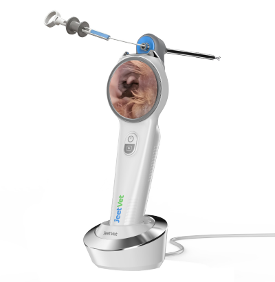
JeetVet Veterinary Endoscopy for Companion Animal Practice
JeetVet develops veterinary endoscope solutions for companion animal and exotic animal applications, with configurations designed for different patient sizes and clinical procedures.
For veterinary practices, selecting an endoscope according to insertion diameter, working length, imaging requirements, and instrument channel needs can help build a more practical endoscopy workflow.
JeetVet focuses on combining clear visualization, maneuverability, portability, and accessory compatibility to support veterinarians in gastrointestinal examination, biopsy, foreign body retrieval, and other minimally invasive applications.
Conclusion
Chronic vomiting in cats can have many possible causes, and identifying the underlying problem may require more than routine examination alone.
When clinically indicated, a veterinary endoscope provides direct visualization of the upper gastrointestinal tract and enables targeted biopsy through a minimally invasive approach.
For veterinary clinics treating feline patients, gastrointestinal endoscopy can be a valuable addition to the diagnostic toolkit—helping clinicians gather more precise information and make better-informed treatment decisions.