Veterinary Endoscope for Chronic Diarrhea in Dogs and Cats: A Practical Guide to Gastrointestinal Examination and Biopsy
Chronic diarrhea is a common clinical concern in dogs and cats. When gastrointestinal symptoms persist or recur despite initial management, veterinarians may need additional diagnostic information to identify the underlying cause and develop an appropriate treatment plan.
A veterinary endoscope allows clinicians to directly examine accessible areas of the gastrointestinal tract and collect tissue samples for histopathological evaluation. In selected patients, veterinary gastrointestinal endoscopy can provide valuable diagnostic information without requiring a surgical approach to obtain mucosal biopsies.
This guide explores how veterinary endoscopy supports the evaluation of chronic diarrhea, when endoscopic biopsy may be considered, and which equipment features matter in clinical practice.
1. Understanding Chronic Diarrhea in Dogs and Cats
Chronic diarrhea generally refers to diarrhea that persists or recurs over several weeks. It may involve the small intestine, large intestine, or both.
Possible underlying causes include:
- Diet-responsive enteropathies and adverse food reactions
- Parasitic or infectious gastrointestinal diseases
- Chronic inflammatory enteropathies
- Intestinal lymphangiectasia and other disorders affecting nutrient absorption
- Gastrointestinal neoplasia, including intestinal lymphoma
- Extraintestinal disorders that affect gastrointestinal function
Clinical signs may include increased stool frequency, weight loss, vomiting, altered appetite, mucus in the stool, or changes in stool consistency.
Because different disorders may produce similar symptoms, a structured diagnostic approach is essential.
2. When Should Veterinary Endoscopy Be Considered?
A veterinary endoscope is not necessarily the first diagnostic tool used for chronic diarrhea. Initial evaluation commonly includes a clinical history, physical examination, fecal testing, laboratory investigations, and appropriate dietary assessment or trials.
Veterinarians may consider gastrointestinal endoscopy when:
- Diarrhea persists despite an appropriate initial diagnostic and management plan.
- There is unexplained weight loss or concern about intestinal malabsorption.
- Clinical findings raise suspicion of chronic inflammatory enteropathy or gastrointestinal neoplasia.
- Histopathological examination is needed to help distinguish possible intestinal disorders.
- Direct assessment of accessible gastrointestinal mucosa may influence clinical decisions.
The decision to perform endoscopy should consider the patient's condition, diagnostic findings, anesthetic risk, and the expected value of tissue sampling.
3. How a Veterinary Endoscope Supports Gastrointestinal Examination
Direct Visualization of the Gastrointestinal Mucosa
Flexible veterinary endoscopes provide real-time visualization of accessible gastrointestinal surfaces. Depending on the examination route and equipment, veterinarians may inspect the stomach, portions of the small intestine, colon, and selected additional intestinal segments.
During the procedure, clinicians may evaluate visible findings such as:
- Mucosal redness or altered coloration
- Erosions and ulceration
- Changes in mucosal texture or appearance
- Abnormal masses or localized lesions
- Other visible changes requiring further investigation
These findings can help guide tissue sampling, although a normal-looking mucosa does not exclude microscopic gastrointestinal disease.
Supporting Targeted Tissue Sampling
When a veterinary endoscope includes a compatible working channel, biopsy forceps can be introduced to collect small mucosal tissue samples under direct visualization.
Endoscopic biopsy may help veterinarians investigate conditions such as chronic inflammatory enteropathy and certain forms of intestinal neoplasia.
However, endoscopic biopsies primarily sample the mucosa. Diseases involving deeper intestinal layers or inaccessible regions may require alternative sampling methods.
4. Veterinary Endoscopic Biopsy: Why Tissue Quality Matters
Obtaining adequate biopsy samples is an important part of diagnostic gastrointestinal endoscopy.
Several factors influence the usefulness of endoscopic biopsies:
- Sampling location: Tissue should be collected from clinically relevant and accessible gastrointestinal regions.
- Sample quantity: Multiple samples may be needed to improve diagnostic assessment, following the veterinarian's sampling protocol.
- Instrument compatibility: Biopsy forceps must be suitable for the endoscope's working channel.
- Tissue handling: Careful collection, preservation, and submission support histopathological interpretation.
- Clinical context: Biopsy results should be interpreted alongside patient history, laboratory findings, and other investigations.
Endoscopy is a method of obtaining diagnostic information rather than a standalone diagnosis. Histopathology and the broader clinical assessment remain essential.
5. Choosing a Veterinary Endoscope for Gastrointestinal Examination
Veterinary clinics evaluating endoscopy equipment should consider more than image resolution alone. Several design features can influence practical usability and examination capability.
| Equipment Feature | Clinical Importance |
|---|---|
| Insertion Tube Diameter | Should be appropriate for patient size and the intended examination route. |
| Working Length | Determines whether the endoscope can reach the intended anatomical region. |
| Working Channel | Allows compatible biopsy forceps and other instruments to be introduced. |
| Image Quality | Supports examination of mucosal surfaces and instrument positioning. |
| Tip Articulation | Helps clinicians navigate anatomical curves and adjust the viewing direction. |
| Cleaning and Reprocessing | Requires validated procedures compatible with the equipment and accessories. |
Clinics treating both dogs and cats may require different endoscope configurations to accommodate differences in anatomy and patient size.
6. Important Limitations of Gastrointestinal Endoscopy
Although veterinary endoscopy offers important diagnostic advantages, it also has limitations.
- Standard gastrointestinal endoscopes cannot examine every part of the small intestine.
- Some intestinal disorders occur beyond the reach of conventional endoscopic equipment.
- Superficial mucosal biopsies may not adequately characterize deeper intestinal disease.
- Endoscopic appearance alone cannot reliably distinguish all inflammatory and neoplastic conditions.
- Procedures generally require anesthesia and appropriate patient monitoring.
In certain cases, additional imaging, laboratory testing, surgical biopsy, or referral to a specialist may be necessary.
7. JeetVet Veterinary Endoscopy Solutions for Clinical Practice
JeetVet develops veterinary endoscopy equipment designed to support visualization and examination across a range of veterinary clinical applications.
For gastrointestinal procedures, equipment selection should account for the animal's size, the intended anatomical region, and the need for accessory instruments such as biopsy forceps.
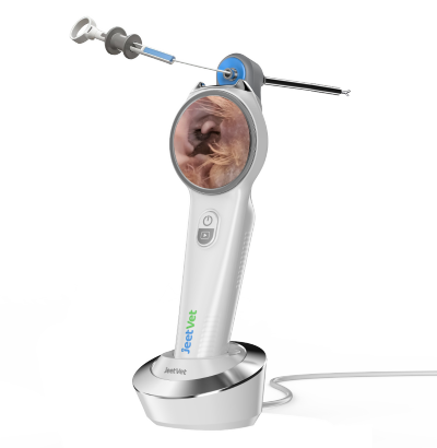
The JeetVet RAE-105 Portable USB Veterinary Endoscope offers a portable examination configuration with a working channel that can support compatible instruments for selected procedures. Its suitability for a particular gastrointestinal examination or biopsy should be confirmed against the required anatomical reach and accessory specifications.
By considering endoscope diameter, working length, imaging capability, and working-channel compatibility, veterinary clinics can identify equipment configurations aligned with their diagnostic workflows.
Explore JeetVet veterinary endoscopy solutions at www.jeetvet.com.
8. Frequently Asked Questions
Can a veterinary endoscope diagnose the cause of chronic diarrhea?
A veterinary endoscope can provide direct visualization and facilitate mucosal biopsy. The underlying diagnosis generally requires interpretation of biopsy findings together with other clinical investigations.
Is veterinary endoscopy suitable for both dogs and cats?
Yes. Gastrointestinal endoscopy is used in both species, but endoscope size, examination route, patient condition, and procedure objectives must be considered individually.
Why is a working channel important for veterinary gastrointestinal endoscopy?
A working channel permits the introduction of compatible instruments, including biopsy forceps, which can be important when tissue sampling is required.
Can a normal endoscopic appearance rule out intestinal disease?
No. Some gastrointestinal disorders cause microscopic changes that are not visible during endoscopy. Biopsy may still be appropriate when supported by the clinical findings.
Can endoscopy replace surgical intestinal biopsy?
Not in every case. Endoscopic biopsy provides mucosal samples from accessible areas, while surgical biopsy may be necessary when deeper tissue or inaccessible intestinal regions require evaluation.
Conclusion
Chronic diarrhea in dogs and cats can present significant diagnostic challenges because many gastrointestinal disorders produce overlapping clinical signs.
A veterinary endoscope can support a structured diagnostic approach by enabling direct mucosal examination and, when appropriate, endoscopic biopsy. Understanding the capabilities and limitations of gastrointestinal endoscopy helps veterinary teams determine when the procedure may provide meaningful clinical information.
For clinics considering veterinary endoscopy equipment, appropriate working length, insertion tube diameter, image quality, and working-channel compatibility are key factors in selecting a suitable solution.