Veterinary Endoscope for Chronic Vomiting in Dogs and Cats: A Practical Guide to Upper Gastrointestinal Examination
Chronic vomiting is a common reason for veterinary consultations in dogs and cats. While occasional vomiting may have a relatively simple explanation, persistent or recurrent episodes can indicate underlying gastrointestinal or systemic disorders that require further investigation.
When initial diagnostic tests do not provide sufficient answers, a veterinary endoscope may help clinicians directly examine accessible areas of the upper gastrointestinal tract and obtain mucosal biopsy samples when indicated.
This guide explains when veterinary upper gastrointestinal endoscopy may be considered, how it supports clinical assessment, and which endoscope features are important for veterinary clinics.
1. Understanding Chronic Vomiting in Dogs and Cats
Chronic vomiting generally refers to vomiting that persists or recurs over an extended period. The frequency, duration, associated clinical signs, and patient history all influence the diagnostic approach.
Potential causes include:
- Dietary intolerance or food-responsive gastrointestinal disorders
- Chronic inflammatory gastrointestinal disease
- Gastric or duodenal ulceration
- Gastrointestinal foreign material or partial obstruction
- Gastrointestinal neoplasia
- Metabolic, endocrine, hepatic, renal, or other systemic disorders
Patients may also show reduced appetite, weight loss, lethargy, abdominal discomfort, or changes in stool consistency.
Because vomiting is not specific to a single disease, veterinarians should assess the patient comprehensively before deciding whether endoscopy is appropriate.
2. When Should Veterinary Gastroscopy Be Considered?
Veterinary gastroscopy is generally considered after an appropriate initial diagnostic assessment, which may include clinical history, physical examination, laboratory testing, fecal examination, diagnostic imaging, and selected dietary or medical management.
A veterinary endoscope may be useful when:
- Vomiting persists despite appropriate initial investigation or management.
- There is concern about gastric or proximal intestinal mucosal disease.
- Direct visualization of the esophagus, stomach, or proximal duodenum could help guide further decisions.
- Gastrointestinal mucosal biopsy is needed for histopathological examination.
- A suspected upper gastrointestinal foreign body may be accessible for endoscopic evaluation.
Endoscopy should not delay urgent stabilization or treatment when a patient has signs of severe illness, suspected perforation, or another emergency condition.
3. How a Veterinary Endoscope Supports Upper Gastrointestinal Examination
Direct Visualization of the Esophagus and Stomach
A flexible veterinary endoscope allows veterinarians to inspect accessible upper gastrointestinal surfaces in real time.
Depending on the procedure and equipment, the examination may include the esophagus, stomach, and proximal duodenum.
Clinicians may identify visible abnormalities such as:
- Mucosal redness or altered appearance
- Erosions and ulceration
- Abnormal tissue growths or masses
- Retained foreign material
- Changes in mucosal texture or surface appearance
Visual findings may help guide further investigation, but normal endoscopic appearance does not exclude microscopic disease.
Supporting Gastrointestinal Biopsy
When the endoscope includes a suitable working channel, compatible biopsy forceps can be introduced to obtain mucosal tissue samples.
Histopathological examination may help investigate chronic inflammatory conditions and selected gastrointestinal neoplasms.
However, standard endoscopic biopsies primarily sample the mucosa. Deeper lesions or disease outside the accessible examination area may require additional diagnostic methods.
4. Why Air, Water, and Suction Functions Matter During Gastroscopy
Upper gastrointestinal endoscopy requires an adequately visible examination field. Depending on the endoscope configuration, air insufflation, water irrigation, and suction can support the procedure.
Air Insufflation
Controlled insufflation helps expand the stomach and separate mucosal folds, allowing more complete inspection of accessible surfaces.
Excessive distension should be avoided, and insufflation should follow appropriate clinical protocols.
Water Irrigation
Water irrigation can help clear mucus and debris from the lens or examination area, improving visualization when supported by the system.
Suction
Suction can remove excess irrigation fluid and selected loose contents, helping maintain visibility during examination.
Not all veterinary endoscopes include these functions, and clinics should verify the configuration of each model before purchase.
5. Choosing the Right Veterinary Endoscope for Gastroscopy
Veterinary clinics should select endoscopy equipment according to the patient population, intended anatomical reach, and diagnostic requirements.
| Equipment Feature | Clinical Importance |
|---|---|
| Insertion Tube Diameter | Should be appropriate for the patient's anatomy and examination route. |
| Working Length | Must be sufficient to reach the intended upper gastrointestinal region. |
| Image Quality | Supports detailed visualization of accessible mucosal surfaces. |
| Working Channel | Allows compatible biopsy forceps and other instruments to be introduced. |
| Tip Articulation | Helps clinicians navigate anatomical curves and adjust viewing direction. |
| Air, Water, and Suction | Supports lumen distension, irrigation, and fluid management when available. |
Clinics treating a wide range of dog and cat sizes may require more than one endoscope configuration.
6. Important Clinical Limitations
Although veterinary gastroscopy can provide useful diagnostic information, it has several limitations.
- It does not evaluate every part of the gastrointestinal tract.
- Some disorders produce microscopic changes without obvious visual abnormalities.
- Standard endoscopic biopsy may not adequately sample deeper gastrointestinal lesions.
- Endoscopy cannot independently diagnose all causes of chronic vomiting.
- Procedures generally require anesthesia, appropriate patient preparation, and monitoring.
In some patients, additional laboratory tests, diagnostic imaging, advanced sampling, or referral may be necessary.
7. JeetVet Veterinary Endoscopy Solutions
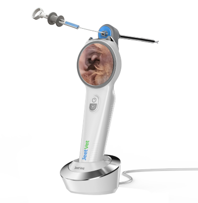
JeetVet develops portable veterinary endoscopy equipment designed to support clinical visualization and selected diagnostic procedures.
The JeetVet RAE-105 Portable USB Veterinary Endoscope provides high-definition imaging, a 1000 mm working length, and a working channel for compatible instruments. Water and air supply functions are available for supported examination workflows.
The RAE-105Pro Portable WiFi Veterinary Endoscope offers wireless connectivity together with water, air, and suction capabilities, providing an alternative configuration for clinics evaluating portable veterinary endoscopy systems.
Equipment suitability should always be confirmed against patient size, required anatomical reach, instrument compatibility, and the specific clinical procedure.
Discover JeetVet veterinary endoscopy solutions at www.jeetvet.com.
8. Frequently Asked Questions
Can a veterinary endoscope help investigate chronic vomiting in dogs?
Yes. In selected patients, upper gastrointestinal endoscopy can help veterinarians inspect accessible mucosal surfaces and collect biopsy samples when clinically indicated.
Can cats undergo veterinary gastroscopy?
Yes. Gastroscopy can be performed in cats using appropriately sized equipment and suitable anesthetic and clinical protocols.
Does veterinary endoscopy always require a biopsy?
No. The need for biopsy depends on the clinical question and examination findings. When microscopic disease is suspected, biopsy may provide important diagnostic information.
Why is a working channel important in veterinary gastroscopy?
A working channel allows compatible instruments, such as biopsy forceps, to be introduced during selected procedures.
Can gastroscopy identify every cause of vomiting?
No. Vomiting may result from gastrointestinal or systemic conditions, and some disorders cannot be identified through gastroscopy alone.
Conclusion
Persistent vomiting in dogs and cats can be associated with a wide range of underlying disorders. A structured diagnostic approach helps veterinarians determine when additional gastrointestinal investigation is appropriate.
In selected cases, a veterinary endoscope supports upper gastrointestinal examination by providing direct visualization and access for mucosal biopsy.
For veterinary clinics, choosing equipment with appropriate imaging capabilities, working length, insertion diameter, working-channel compatibility, and fluid-management functions can support practical endoscopy workflows.