Veterinary Endoscope for Otoscopy: A Practical Guide to Ear Examination in Dogs and Cats
Ear problems are among the common reasons dogs and cats visit veterinary clinics. From persistent scratching and head shaking to ear discharge, unpleasant odors, and recurrent inflammation, ear conditions can affect an animal's comfort and quality of life.
While conventional otoscopic examination is useful for evaluating the external ear canal, some clinical situations require more detailed visualization. A veterinary endoscope can help veterinarians examine the ear canal, assess visible abnormalities, and support selected ear cleaning and irrigation procedures.
Veterinary otoscopy is particularly valuable when investigating chronic otitis externa, suspected ear canal foreign bodies, abnormal tissue changes, and recurrent ear infections.
This guide explains how veterinary endoscopy supports ear examination in dogs and cats, what veterinarians should consider during otoscopy, and how to select appropriate veterinary ear examination equipment.
1. What Is Veterinary Otoscopy?
Veterinary otoscopy is the examination of an animal's external ear canal and accessible ear structures using an appropriate viewing instrument.
A veterinary endoscope or specialized veterinary otoscope can provide illuminated visualization, helping clinicians assess areas that may be difficult to examine through a routine external inspection.
Depending on the equipment and examination approach, veterinary otoscopy may help identify:
- Excessive earwax accumulation.
- Redness and inflammation of the ear canal.
- Ear canal discharge and debris.
- Foreign materials within the ear canal.
- Abnormal tissue growths or masses.
- Changes in the visible tympanic membrane.
Direct visualization is important because different ear conditions may produce similar external symptoms but require different diagnostic and treatment approaches.
2. Common Ear Problems in Dogs and Cats
Dogs and cats may develop several conditions affecting the external ear canal. Understanding the clinical presentation helps veterinarians determine whether further endoscopic examination is necessary.
Otitis Externa
Otitis externa refers to inflammation of the external ear canal. It is commonly associated with symptoms such as:
- Frequent head shaking.
- Scratching around the ears.
- Redness and swelling.
- Unusual ear discharge.
- Unpleasant odor.
- Sensitivity when the ears are touched.
Veterinary otoscopy allows clinicians to evaluate visible ear canal changes and assess whether debris or inflammation is interfering with examination.
Earwax and Debris Accumulation
Excessive cerumen, commonly known as earwax, can obstruct visualization of the ear canal.
In selected cases, appropriate ear cleaning or irrigation may help remove accumulated material and improve visibility.
Ear Canal Foreign Bodies
Plant material, grass awns, and other foreign objects may enter the ear canal and cause irritation, pain, and inflammation.
Direct endoscopic visualization can help identify the location and characteristics of accessible foreign materials.
Chronic or Recurrent Ear Disease
Some animals experience repeated episodes of ear inflammation despite previous treatment.
Further examination may be necessary to investigate persistent abnormalities, evaluate the ear canal, and identify possible underlying conditions.
3. How a Veterinary Endoscope Improves Ear Examination
A veterinary endoscope provides direct visualization of accessible ear canal structures. This can help veterinarians assess abnormalities more systematically.
Clear Visualization of the Ear Canal
Illumination and magnified imaging can help clinicians examine the ear canal surface and identify visible changes such as redness, swelling, discharge, or abnormal tissue.
Assessment of Ear Canal Obstruction
Earwax, inflammatory debris, and foreign materials may partially or completely obstruct the examination field.
Endoscopic visualization can help determine the extent and location of visible obstruction.
Evaluation of the Tympanic Membrane
When the ear canal is sufficiently accessible and visibility is adequate, veterinarians may examine the tympanic membrane, also known as the eardrum.
Assessing its appearance is important when investigating certain ear conditions and before performing procedures involving ear irrigation.
Supporting Ear Cleaning Procedures
Depending on the equipment configuration, veterinary endoscopy may support controlled ear cleaning and irrigation while allowing clinicians to monitor the accessible examination area.
The procedure should always be performed according to the patient's condition, anatomical findings, and appropriate clinical protocols.
4. Veterinary Ear Canal Irrigation: Why Visualization Matters
Ear canal irrigation is used in selected clinical situations to help remove accumulated debris, cerumen, and other material from the external ear canal.
However, irrigation should not be performed automatically in every patient with ear discharge or obstruction.
Before selecting an irrigation approach, veterinarians should evaluate:
- The degree of ear canal inflammation.
- The amount and characteristics of accumulated material.
- The accessibility of the ear canal.
- The condition of the tympanic membrane.
- Whether tympanic membrane integrity can be established.
- Potential contraindications to irrigation.
Direct visualization can help veterinarians monitor the cleaning process and assess visible changes within the accessible ear canal.
When tympanic membrane integrity is uncertain or the ear canal is severely diseased, additional caution and an appropriate specialist-directed approach are necessary.
5. The Role of Veterinary Endoscopy in Ear Foreign Body Examination
Foreign bodies within the ear canal can cause sudden discomfort, persistent head shaking, scratching, and localized inflammation.
Common examples include:
- Grass awns.
- Small plant fragments.
- Seeds and other organic materials.
- Small pieces of debris.
A veterinary endoscope can help identify accessible foreign material and provide a clearer view of its position.
When removal is appropriate, compatible instruments may be used under direct visualization if the equipment supports the procedure.
Important considerations include:
- Foreign body size and shape.
- Its position within the ear canal.
- Whether it is embedded in tissue.
- The degree of inflammation or bleeding.
- The risk of damaging the ear canal or tympanic membrane.
Not every ear canal foreign body can be removed with an endoscope. Some cases require specialized instruments, anesthesia, referral, or an alternative treatment approach.
6. Why Image Quality Matters in Veterinary Otoscopy
The ear canal is a narrow anatomical structure, and small visible abnormalities may be clinically important.
A veterinary endoscope should provide suitable imaging performance to support detailed examination.
Important imaging considerations include:
- Resolution: Helps visualize small details on the ear canal surface.
- Illumination: Provides sufficient light in narrow examination spaces.
- Color reproduction: Supports observation of visible tissue coloration.
- Image stability: Helps maintain a consistent view during examination.
- Viewing angle: Influences the anatomical area that can be observed.
Resolution alone does not determine clinical performance. The complete imaging system, illumination, viewing distance, and examination technique all contribute to the quality of visualization.
7. Choosing the Right Veterinary Endoscope for Ear Examination
Veterinary clinics should consider several equipment characteristics when selecting an endoscope for otoscopy.
| Equipment Feature | Clinical Importance |
|---|---|
| Insertion Diameter | Must be appropriate for the patient's ear canal dimensions. |
| Image Resolution | Supports detailed visualization of accessible ear structures. |
| Illumination | Provides adequate visibility in narrow anatomical spaces. |
| Viewing Angle | Influences examination coverage and visibility. |
| Irrigation Capability | May support selected ear cleaning procedures when clinically appropriate. |
| Handling and Control | Supports controlled examination and instrument positioning. |
| Cleaning and Reprocessing | Must comply with the manufacturer's instructions for the specific device. |
Clinics should also consider whether the system is intended specifically for ear examination or can support multiple veterinary endoscopy applications.
8. Veterinary Otoscope vs. Veterinary Endoscope
Although the terms are sometimes used interchangeably, veterinary otoscopes and veterinary endoscopes can refer to different equipment configurations.
| Comparison | Veterinary Otoscope | Veterinary Endoscope |
|---|---|---|
| Primary Application | External ear examination | Multiple diagnostic and procedural applications depending on configuration |
| Visualization | Viewing the ear canal and visible tympanic membrane | Direct visualization of accessible internal structures |
| Accessories | Depends on the otoscope design | May support compatible biopsy or retrieval instruments |
| Clinical Flexibility | Primarily ear examination | Depends on the endoscope model and accessories |
The appropriate choice depends on the intended clinical applications, the anatomical structures being examined, and the equipment configuration.
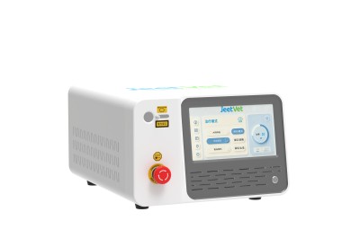
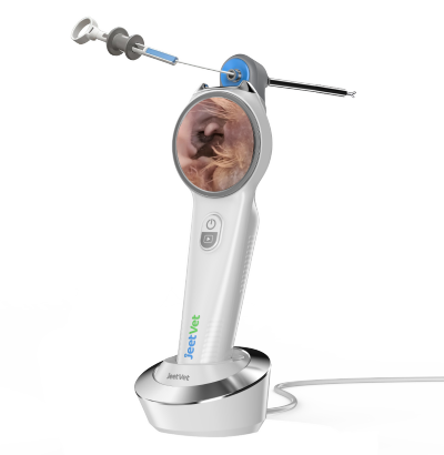
9. JeetVet VET-600: Supporting Veterinary Ear Examination
JeetVet develops veterinary endoscopy solutions designed to support visualization and clinical examination in animal healthcare.
The VET-600 Veterinary Endoscope is designed to support veterinary examination applications, including ear canal visualization and related clinical workflows.
Its fully immersible design is one of its key product features, subject to the specified immersion conditions and manufacturer instructions.
For veterinary clinics, important considerations when selecting an ear examination system include:
- Clear visualization of the ear canal.
- Appropriate examination accessories.
- Compatibility with intended ear cleaning procedures.
- Ease of operation during routine examinations.
- Cleaning and reprocessing requirements.
JeetVet aims to provide practical veterinary endoscopy solutions that help clinicians examine animal health conditions with greater visualization and procedural confidence.
Discover JeetVet veterinary endoscopy products at www.jeetvet.com.
10. Frequently Asked Questions
What is veterinary otoscopy used for?
Veterinary otoscopy is used to examine the external ear canal and accessible ear structures, helping veterinarians identify inflammation, accumulated debris, foreign materials, and other visible abnormalities.
Can a veterinary endoscope help examine chronic ear infections?
Yes. Endoscopic visualization may help assess accessible ear canal surfaces, identify visible abnormalities, and support further diagnostic evaluation in animals with recurrent ear disease.
Can veterinary endoscopy help remove ear canal foreign bodies?
In selected cases, an appropriately configured endoscope and compatible instruments may support foreign body retrieval under direct visualization.
Can an endoscope be used for ear canal irrigation?
Some veterinary endoscopy systems support irrigation or are compatible with appropriate irrigation accessories. The procedure should only be performed when clinically appropriate and with consideration of tympanic membrane integrity.
Why is the tympanic membrane important during ear examination?
The tympanic membrane separates the external ear canal from the middle ear. Its condition is an important consideration when evaluating ear disease and determining whether irrigation or topical treatments are appropriate.
What should clinics consider when purchasing a veterinary otoscope?
Important considerations include imaging performance, insertion diameter, illumination, examination accessories, irrigation compatibility, cleaning requirements, and the clinical applications the equipment is designed to support.
Conclusion
Ear conditions in dogs and cats can involve inflammation, excessive debris, foreign bodies, and other abnormalities that require careful examination.
A veterinary endoscope can provide direct visualization of accessible ear canal structures and support selected diagnostic and cleaning procedures.
For veterinary clinics, selecting appropriate ear examination equipment requires consideration of insertion diameter, imaging quality, illumination, handling characteristics, and cleaning compatibility.
With suitable equipment and clinical expertise, veterinary otoscopy can become an important part of routine ear examination and the evaluation of recurrent ear disease.