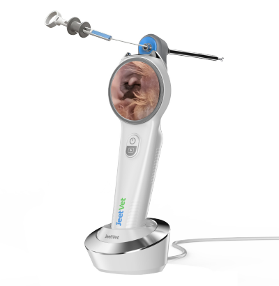
Veterinary Endoscope for Rhinoscopy: A Practical Guide to Nasal Examination in Dogs and Cats
Persistent sneezing, nasal discharge, nosebleeds, and abnormal breathing are common reasons for dogs and cats to be presented for veterinary examination. While some nasal conditions respond to routine treatment, persistent or recurrent clinical signs may require a closer investigation of the nasal cavity.
A veterinary endoscope can provide direct visualization of accessible nasal structures, allowing veterinarians to inspect the mucosal surface, identify foreign material or abnormal tissue, and obtain samples when clinically appropriate.
This procedure, commonly known as veterinary rhinoscopy, has become an important minimally invasive diagnostic technique for investigating selected nasal and upper respiratory conditions in companion animals.
What Is Veterinary Rhinoscopy?
Veterinary rhinoscopy is an endoscopic procedure used to examine the nasal passages and related structures.
Depending on the patient, anatomy, equipment, and clinical objective, veterinarians may use appropriate rigid or flexible endoscopic techniques to investigate the nasal cavity.
A veterinary endoscope provides real-time visualization, enabling the clinician to evaluate structures that cannot be adequately inspected during a routine external examination.
Rhinoscopy may form part of the diagnostic workup for:
- Persistent nasal discharge
- Chronic sneezing
- Recurrent nosebleeds
- Suspected nasal foreign bodies
- Abnormal respiratory noise
- Suspected nasal masses or proliferative tissue
- Chronic nasal inflammation
Why Are Nasal Diseases Difficult to Diagnose?
The nasal cavity has complex internal anatomy and cannot be fully assessed through external inspection alone.
In addition, several different conditions may produce similar clinical signs. Persistent nasal discharge, for example, may be associated with inflammation, foreign material, infection, dental-related disease, fungal disease, or neoplasia.
For this reason, a complete diagnostic approach may involve multiple methods selected according to the individual patient.
When direct visualization is clinically indicated, a veterinary endoscope can provide additional information about the appearance and location of abnormalities within accessible areas of the nasal cavity.
Clinical Signs That May Lead to Rhinoscopy
Veterinarians may consider further nasal investigation when a patient presents with persistent or unexplained signs such as:
- Unilateral or bilateral nasal discharge
- Bloody nasal discharge or epistaxis
- Frequent sneezing
- Nasal discomfort
- Pawing at the nose
- Abnormal respiratory sounds
- Reduced airflow through one nostril
- Visible facial or nasal abnormalities
These signs do not automatically mean that endoscopy is required. The decision depends on the patient's history, physical examination, previous diagnostic findings, and the veterinarian's clinical judgment.
What Can a Veterinary Endoscope Reveal During Rhinoscopy?
Direct visualization can help veterinarians assess the appearance of the nasal mucosa and identify abnormalities in accessible regions.
Possible endoscopic findings may include:
- Mucosal redness or inflammation
- Abnormal secretions
- Localized bleeding
- Foreign material
- Irregular or proliferative tissue
- Changes in normal anatomical structures
Importantly, the appearance of a lesion alone may not establish a definitive diagnosis. Abnormal findings often need to be interpreted together with other clinical information and, when appropriate, laboratory or pathological analysis.
Nasal Foreign Body Detection and Removal
Foreign material is one reason rhinoscopy may be performed, particularly in animals with sudden-onset sneezing, nasal irritation, or unilateral discharge.
Potential nasal foreign bodies may include:
- Grass awns
- Plant material
- Small seeds
- Other environmental debris
When a foreign body is accessible and endoscopic removal is considered appropriate, direct visualization can help the veterinarian identify its location and evaluate the surrounding tissue.
Depending on the endoscope configuration, compatible grasping instruments may then be used to assist retrieval.
Rhinoscopy and Targeted Biopsy
If abnormal tissue is identified during nasal examination, visual appearance alone may not be sufficient to determine its cause.
When clinically appropriate and technically feasible, targeted tissue sampling can help provide additional diagnostic information.
This is where a veterinary endoscope with a suitable working channel can offer additional clinical value. Compatible biopsy instruments may be introduced while the clinician maintains visualization of the target area.
Collected tissue can then be submitted for histopathological evaluation as part of the diagnostic process.
Why Image Quality Matters in Veterinary Rhinoscopy
The nasal cavity contains narrow passages and complex anatomical structures. Clear visualization is therefore particularly important during rhinoscopy.
Clinically useful imaging depends on more than resolution alone. Veterinarians should also consider:
- Illumination quality
- Color reproduction
- Image stability
- Working distance
- Field of view
- Distal tip maneuverability
These factors work together to help the clinician distinguish normal anatomy from areas that may require closer examination.
Choosing a Veterinary Endoscope for Nasal Examination
Not every veterinary endoscope is suitable for every patient or nasal procedure.
When evaluating equipment for rhinoscopy, veterinary clinics should consider:
| Factor | Why It Matters |
|---|---|
| Insertion Diameter | Must be appropriate for patient size and target anatomy. |
| Image Quality | Supports visualization of mucosa and abnormal tissue. |
| Illumination | Provides visibility inside narrow, dark anatomical spaces. |
| Maneuverability | Helps navigate complex anatomical structures. |
| Working Channel | May support compatible biopsy or retrieval instruments. |
| Documentation | Allows images or video to be recorded for clinical review when required. |
Flexible vs. Rigid Endoscopy for Rhinoscopy
Both flexible and rigid endoscopic approaches can have roles in veterinary nasal examination. The appropriate choice depends on patient anatomy, the area being examined, the procedure being performed, and clinician preference.
Flexible endoscopes can provide useful maneuverability through certain anatomical pathways, while appropriately selected rigid endoscopes may provide advantages for other nasal examination techniques.
Rather than assuming that one design is universally better, clinics should choose equipment according to the intended clinical application.
The Role of Veterinary Endoscopy in a Complete Diagnostic Workflow
Rhinoscopy should not be considered an isolated diagnostic solution.
Depending on the patient, veterinarians may combine endoscopic findings with:
- Clinical history
- Physical examination
- Laboratory testing
- Appropriate diagnostic imaging
- Cytology
- Histopathology
- Microbiological testing when indicated
The value of the veterinary endoscope is its ability to add direct visual information and, in selected cases, facilitate targeted sampling or minimally invasive intervention.
How JeetVet Supports Veterinary Endoscopic Examination
JeetVet develops veterinary endoscope solutions for companion animal and exotic animal applications, with configurations designed for different patient sizes and clinical requirements.
For selected respiratory and nasal applications, factors such as insertion diameter, visualization, maneuverability, and instrument compatibility are important when choosing an appropriate endoscopy solution.
JeetVet focuses on combining practical imaging performance, flexible operation, portability, and clinical usability to support veterinarians across a range of minimally invasive examinations.
Conclusion
Persistent nasal signs in dogs and cats can have many possible causes, and direct visualization may provide valuable information when routine examination alone is insufficient.
A veterinary endoscope used for rhinoscopy can help veterinarians examine accessible nasal structures, identify foreign material or abnormal tissue, and support targeted sampling when clinically appropriate.
For veterinary practices developing advanced respiratory diagnostic services, rhinoscopy represents an important application of modern veterinary endoscopy and can contribute to a more complete, minimally invasive diagnostic workflow.