Veterinary Endoscope for Rhinoscopy: A Practical Guide to Nasal Examination in Dogs and Cats
Nasal disorders are common reasons for veterinary consultations in dogs and cats. Persistent sneezing, nasal discharge, nosebleeds, and noisy breathing may indicate underlying conditions that require further investigation.
While physical examination and diagnostic imaging provide important information, some nasal conditions require direct visualization of the nasal passages. A veterinary endoscope can help veterinarians examine accessible nasal structures, identify abnormalities, and collect tissue samples when appropriate.
Veterinary rhinoscopy is particularly valuable when investigating suspected nasal foreign bodies, chronic inflammation, abnormal tissue growth, and unexplained nasal bleeding.
This guide explains how veterinary endoscopy supports nasal examination, which clinical conditions may benefit from rhinoscopy, and what equipment features veterinary clinics should consider.
1. What Is Veterinary Rhinoscopy?
Veterinary rhinoscopy is an endoscopic examination of the nasal passages and associated structures in animals.
Using an appropriately sized veterinary endoscope, clinicians can directly visualize accessible nasal surfaces and investigate abnormalities that may not be fully characterized through external examination alone.
Depending on the equipment and examination approach, rhinoscopy may help veterinarians:
- Examine the nasal cavity.
- Identify visible foreign materials.
- Evaluate abnormal mucosal changes.
- Investigate suspected nasal masses.
- Identify areas of inflammation or bleeding.
- Collect targeted biopsy samples using compatible instruments.
Rhinoscopy may be performed through different approaches depending on the suspected location of the disease and the patient's anatomy.
2. When Should Dogs and Cats Undergo Nasal Endoscopy?
Several clinical signs may indicate the need for further nasal investigation.
Persistent Sneezing
Frequent or persistent sneezing may be associated with nasal irritation, foreign material, inflammation, infection, or other nasal disorders.
When initial investigations do not identify the cause, rhinoscopy may provide additional information through direct examination.
Chronic Nasal Discharge
Long-lasting nasal discharge may occur with inflammatory disease, infection, fungal disease, foreign bodies, or nasal masses.
Endoscopic examination can help identify visible abnormalities and guide further diagnostic sampling.
Unexplained Nosebleeds
Recurrent nasal bleeding, also known as epistaxis, requires careful evaluation.
A veterinary endoscope may help identify visible bleeding sites, abnormal tissue, or other findings within accessible nasal passages.
Suspected Nasal Foreign Bodies
Grass awns, plant material, and other small objects may enter the nasal passages and cause irritation, sneezing, or discharge.
In selected cases, rhinoscopy allows veterinarians to locate the foreign material and assess whether endoscopic retrieval is appropriate.
Suspected Nasal Masses
Abnormal tissue growth within the nasal cavity may cause progressive respiratory symptoms, discharge, or bleeding.
Direct visualization and targeted biopsy may help support further diagnosis, although imaging and histopathology are often necessary.
3. How a Veterinary Endoscope Supports Nasal Examination
The nasal cavity contains narrow anatomical pathways and delicate mucosal surfaces. Direct visualization can provide valuable information when investigating suspected nasal disease.
During rhinoscopy, an appropriately selected veterinary endoscope may support several diagnostic tasks.
Direct Visualization
The endoscope provides an illuminated view of accessible nasal structures, allowing the veterinarian to inspect the mucosal surface and identify visible abnormalities.
Foreign Body Identification
Direct visualization may help locate plant material or other foreign objects within the accessible nasal passages.
If retrieval is considered appropriate, compatible instruments may be used under endoscopic guidance when the scope configuration permits.
Targeted Biopsy
When abnormal tissue is identified, compatible biopsy instruments may help obtain samples for histopathological examination.
Biopsy results can provide additional information when investigating inflammatory conditions, fungal disease, and suspected neoplasia.
Post-Procedure Assessment
Following an intervention, endoscopic visualization may help assess accessible areas for remaining foreign material or visible tissue changes.
4. Why Insertion Diameter Matters in Veterinary Rhinoscopy
One of the most important considerations when selecting a veterinary endoscope for rhinoscopy is insertion tube diameter.
The nasal passages are relatively narrow, and their dimensions vary between species and individual animals.
An endoscope with an unsuitable outer diameter may limit access or make examination difficult.
When selecting equipment, veterinary clinics should consider:
- Animal species and body size.
- Diameter of the intended anatomical passage.
- Required viewing direction.
- Whether biopsy or retrieval instruments are needed.
- Flexibility and maneuverability.
- The intended examination route.
Smaller-diameter instruments may be particularly useful when examining narrow anatomical spaces. However, the smallest available diameter is not automatically the best choice. Imaging capability, working channel requirements, and procedural suitability must also be considered.
5. Flexible vs. Rigid Veterinary Endoscopes for Nasal Examination
Veterinary rhinoscopy may require different equipment configurations depending on the anatomical target and clinical objective.
Flexible Veterinary Endoscopes
A flexible veterinary endoscope can navigate curved anatomical pathways and may be useful when examination requires directional movement.
Important considerations include:
- Insertion tube flexibility.
- Distal tip articulation.
- Insertion diameter.
- Working length.
- Image quality.
- Working channel availability.
Ultra-Thin Rigid Endoscopes
Ultra-thin rigid endoscopes may be suitable for selected examinations involving narrow anatomical spaces where a rigid approach is appropriate.
Their potential advantages include:
- Very small outer diameter in selected configurations.
- Stable insertion characteristics.
- Direct visualization of accessible anatomical structures.
- Suitability for certain specialized examination requirements.
Rigid and flexible systems are not interchangeable in every clinical situation. The appropriate choice depends on anatomy, examination route, patient size, and procedural requirements.
6. Image Quality: Seeing Small Nasal Abnormalities Clearly
Clear imaging is essential when examining narrow anatomical spaces.
Veterinarians may need to identify subtle mucosal changes, small foreign materials, bleeding points, or abnormal tissue surfaces.
Important imaging considerations include:
- Resolution: Supports visualization of small anatomical details.
- Illumination: Provides sufficient light within narrow passages.
- Color reproduction: Helps distinguish visible tissue characteristics.
- Field of view: Determines how much of the examination area can be observed.
- Image stability: Supports consistent observation during instrument movement.
High resolution alone does not guarantee effective clinical visualization. Imaging performance should be evaluated together with illumination, viewing distance, and the complete endoscope design.
7. The Role of Veterinary Endoscopy in Nasal Foreign Body Removal
Nasal foreign bodies are an important consideration in dogs and cats presenting with sudden sneezing, nasal irritation, or unilateral nasal discharge.
Plant material and other foreign objects may become lodged within accessible nasal passages.
When an object is identified, the veterinarian must assess:
- Its exact location.
- Its size and shape.
- Whether it is embedded in surrounding tissue.
- The possibility of bleeding or tissue injury.
- Whether compatible retrieval instruments can reach it.
In selected cases, endoscopic guidance can help support controlled retrieval while allowing the veterinarian to observe the surrounding tissues.
Not every nasal foreign body is suitable for endoscopic removal. Some cases require additional imaging, alternative instruments, or surgical intervention.
8. Rhinoscopy and Nasal Biopsy
When a veterinarian identifies suspicious tissue during nasal examination, biopsy may be recommended to investigate the underlying condition.
Histopathological evaluation can help distinguish between different types of inflammatory, infectious, and neoplastic conditions.
Important considerations include:
- Choosing an appropriate sampling location.
- Using compatible biopsy instruments.
- Obtaining sufficient tissue when clinically appropriate.
- Managing bleeding risk.
- Preserving samples according to laboratory requirements.
Because nasal tissues can be vascular and delicate, biopsy procedures require careful clinical judgment and appropriate patient monitoring.
9. How to Choose a Veterinary Endoscope for Rhinoscopy
Before purchasing a veterinary endoscope for nasal examination, clinics should evaluate the complete equipment configuration.
| Feature | Why It Matters |
|---|---|
| Insertion Diameter | Must be appropriate for the narrow anatomical pathway. |
| Image Quality | Supports identification of small visible abnormalities. |
| Illumination | Provides adequate visibility inside the nasal cavity. |
| Flexibility | Helps navigate curved anatomical pathways when required. |
| Working Channel | Allows compatible biopsy or retrieval instruments to be introduced. |
| Working Length | Determines the available reach for the examination. |
| Reprocessing | Must meet the manufacturer's cleaning and disinfection requirements. |
For clinics treating different animal species, selecting equipment based on specific clinical applications is more practical than assuming one endoscope configuration can meet every requirement.
10. JeetVet Solutions for Veterinary Endoscopy
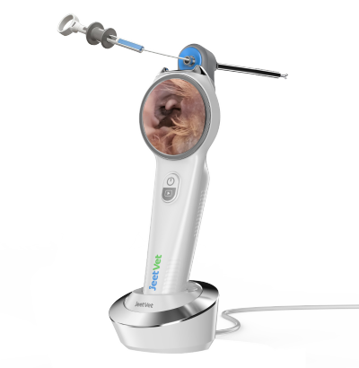
JeetVet develops veterinary endoscopy equipment designed for different animal sizes and clinical examination requirements.
Its product range includes flexible veterinary endoscopes and ultra-thin rigid endoscope configurations for selected diagnostic applications.
The DZT-1616 Ultra-Thin Rigid Endoscope features a 1.6 mm outer diameter and is designed for specialized examination requirements involving narrow anatomical spaces. Its application suitability should be evaluated according to the intended examination route and clinical requirements.
JeetVet also offers portable flexible veterinary endoscopy solutions, including the RAE-105, for appropriate gastrointestinal and other compatible clinical examinations.
When selecting a veterinary endoscope for rhinoscopy, veterinarians should consider insertion diameter, anatomical access, imaging performance, maneuverability, and instrument compatibility together.
Discover JeetVet veterinary endoscopy solutions at www.jeetvet.com.
11. Frequently Asked Questions
What is veterinary rhinoscopy?
Veterinary rhinoscopy is an endoscopic examination of the nasal passages that allows veterinarians to visualize accessible nasal structures and investigate abnormalities.
Can a veterinary endoscope detect nasal foreign bodies?
Yes. An appropriately selected endoscope can help identify visible foreign materials within accessible nasal passages.
Can nasal endoscopy be used to investigate nosebleeds in dogs?
Rhinoscopy may help identify visible abnormalities or potential bleeding sites, although additional diagnostic tests may be required to determine the underlying cause.
Is a flexible or rigid veterinary endoscope better for rhinoscopy?
Neither is universally better. Flexible scopes may be useful for navigating curved passages, while ultra-thin rigid scopes may be appropriate for selected accessible anatomical regions.
What diameter veterinary endoscope is suitable for nasal examination?
The appropriate diameter depends on the animal's size, anatomical passage, examination route, and whether instruments are required. Equipment specifications should be checked before selecting a model.
Can veterinary rhinoscopy replace CT imaging?
No. Rhinoscopy provides direct visualization of accessible nasal surfaces, while CT can evaluate deeper anatomical structures and the extent of disease. The two methods may complement each other.
Conclusion
Nasal disorders in dogs and cats can have multiple underlying causes, ranging from foreign bodies and inflammation to fungal disease and neoplasia.
A veterinary endoscope provides direct visualization of accessible nasal passages and may support foreign body identification, targeted biopsy, and further clinical investigation.
Choosing the appropriate equipment requires careful consideration of insertion diameter, image quality, illumination, flexibility, and accessory compatibility.
With suitable equipment and clinical expertise, veterinary rhinoscopy can become a valuable part of a comprehensive diagnostic approach to nasal disease.